Header Logo

Pulmonary & Sleep Services

Home / Specialities / Pulmonary & Sleep Services / Interventional Pulmonology

Interventional Pulmonology


Call us Call us on 022-61305000


Interventional Pulmonology and Management of Advanced Lung Diseases -- Beyond The Conventional


Interventional pulmonology is a specialized sub specialty of Pulmonary Medicine using minimally invasive and percutaneous techniques for advanced diagnostic and therapeutic management of various lung disorders. Interventional pulmonology has evolved dramatically over the years, was initially used only to examine and sample the central airways, but has now broadened widely to not just diagnose but even treat various lung and pleura related diseases.

The primary application of interventional pulmonology is in the diagnosis, staging and palliative treatment of patients with lung cancer. Dilatation of tracheo-bronchial strictures, stenting of airways, removal of foreign body, management of unclassified pleural disorders and temporary percutaneous tracheotomies for chronic airway management also fall under the realm of interventional pulmonology.

The Department of Interventional Pulmonology (IP) has been developed specifically to cater to the evolving need of minimally invasive procedures for the diagnosis of infective and non-infective lung disorders, and the staging and palliative treatment of advanced lung cancer.

The department is well equipped with state-of-the-art bronchoscopy suite with most advanced diagnostic and therapeutic technologies required for performing these procedures, and early diagnosis and management of advanced lung diseases including lung cancer.

Services provided by the department of IP:

  • Flexible Bronchoscopy

    Broncho alveolar lavage (BAL), Endobronchial lung biopsy (EBLB), Transbronchial lung biopsy (TBLB), Brush biopsy, Cryobiopsy

    • Flexible bronchoscopy:  Bronchoscopy is the most common interventional pulmonology procedure. During bronchoscopy, a doctor advances a flexible endoscope (bronchoscope) through a person's mouth or nose into the windpipe. The doctor advances the bronchoscope through the airways in each lung, to look for any abnormality and patency of the airways. Images from inside the lung are displayed on a video screen.
      The bronchoscope has a channel at its tip, through which a doctor can pass small tools. Using these tools, the doctor can perform several other interventional pulmonology procedures.
    • Bronchoalveolar lavage (BAL): Bronchoalveolar lavage is performed during bronchoscopy. Sterile water is injected through the bronchoscope into a segment of the lung. The fluid is then suctioned back and sent for tests. BAL helps in diagnosis of infection, cancer, bleeding, and other conditions.
    • Endobronchial lung biopsy (EBLB): During bronchoscopy, a doctor may collect a small piece of tissue from small part of abnormal bronchial mucosa or growth in the airways to diagnose various conditions like cancer, tuberculosis or sarcoidosis.
    • Transbronchial lung biopsy (TBLB): A biopsy from the periphery of lung is taken to diagnose interstitial lung disease or any other diffuse lung disease.
    • Brush biopsy: Used to take sample from abnormal airway mucosa.
    • Cryobiopsy: Used to take biopsy from peripheral lung tissue for diffuse lung disease and has the advantage of getting a bigger tissue size for examination.

    Endobronchial ultrasound (EBUS) –Convex and Radial

    Endobronchial Ultrasound (EBUS) is a technique that uses ultrasound along with bronchoscope to visualize airway wall and structures adjacent to it.

    TYPES OF EBUS:

    There are two forms of EBUS; radial and linear (convex).

    • Radial Probe-EBUS
      The system has ultrasound processor and balloon catheter that are attached to the probe. The balloon is fixed at the tip of the probe. It helps to take biopsies from the peripheral parts of lung which are otherwise not accessible by routine flexible bronchoscopy.
    • Convex Probe-EBUS
      In contrast to the radial probe, convex probe has the advantage of accessing central lesions that are in or adjacent to the lung (mediastinum). High-resolution, real-time ultrasound imaging enables direct visualization of the needle as it penetrates the lymph node which optimises the biopsy sample and makes the procedure relatively safe.

    INDICATIONS OF EBUS:

    • Assess the extent of airway invasion
      EBUS has extended the vision beyond the tracheobronchial wall. With EBUS, the delicate multilayer structure of the tracheobronchial wall can be analyzed. This knowledge becomes decisive for the management of early cancer in the central airways.
    • Peripheral intrapulmonary lesions
      Radial probe EBUS can be used to localize peripheral pulmonary nodules and sampling of the lesion can be done without fluoroscopy.
    • Analysis of mediastinal lesions
      Assessment of mediastinal lymph nodes is important for lung cancer staging and planning appropriate treatment strategy. Once target lymph node is identified, linear probe EBUS allows real-time ultrasound guidance during needle insertion. EBUS-TBNA can be used in the evaluation of mediastinal adenopathy due to other aetiologies like sarcoidosis and tuberculosis.
    • Guidance of endo-bronchial therapy
      EBUS provides useful additional information during various interventions including resection of endo-bronchial lesion, stricture dilatation, airway stenting, laser therapy and argon plasma coagulation.

    CONTRAINDICATIONS

    • Life-threatening cardiac arrhythmias
    • Current or recent myocardial ischemia
    • Poorly controlled heart failure
    • Severe hypoxemia
    • Uncooperative patient

    Additional contraindications to EBUS-TBNA are related to bleeding risk and include following:

    • Current anti-platelet agents (such as Ecosprin, Clopidogrel)
    • Current anticoagulant therapy (such as warfarin)
    • Coagulopathy
    • Thrombocytopenia
    • Elevated blood urea nitrogen or serum creatinine

    COMPLICATIONS

    EBUS and EBUS-TBNA are usually safe procedures. Reported complications are agitation, cough, hypoxia, laryngeal injury, fever, bacteraemia and infection, bleeding, pneumothorax and broken equipment becoming stuck in the airway.

    Medical Thoracoscopy/Pleuroscopy

    Medical Thoracoscopy is a minimally invasive procedure that allows access to the pleural space (space between chest wall and lungs), using a combination of viewing and working instruments. It has become the second most important endoscopic procedure in respiratory medicine after bronchoscopy.

    EQUIPMENT:

    • Rigid Pleuroscope
    • Semi-rigid Pleuroscope: Similar to video bronchoscope

    INDICATIONS:

    • Work-up and diagnosis of indeterminate pleural fluid (effusion)
    • Staging of Lung cancer
    • Site-directed biopsy of parietal pleura
    • Staging of mesothelioma (pleural malignancy)

    CONTRAINDICATIONS:

    1. Absolute:

    • Lack of pleural space

    2. Relative:

    • Refractory cough
    • Severe hypoxemia (low O2 saturation)
    • Coagulopathy including low platelet counts
    • Unstable comorbidities or hemodynamic status
    • Pulmonary arterial hypertension

    COMPLICATIONS:

    • Bleeding after a parietal pleura biopsy
    • Lung perforation and air leak
    • Infection in the pleural space

    PREPARATIONS:

    • Fasting for 6-8 hours
    • Stop blood thinners 3-5 days before the procedure (to confirm with the doctor)
    • Patient has to be in the hospital for 3-5 days

    Rigid bronchoscopy

    In rigid bronchoscopy, a long metal tube (rigid bronchoscope) is advanced into a person’s windpipe and main airways. The rigid bronchoscope’s large diameter allows the doctor to use more sophisticated surgical tools and techniques. Rigid bronchoscopy requires general anesthesia (unconsciousness with assisted breathing), like a surgical procedure. It is imperative to undergo evaluation by the physician as well as anesthesiologist prior to the procedure, which allows discussion of risks and benefits and correction of any reversible contraindication. After general anesthesia is administered, the patient is intubated with the rigid bronchoscope and attached to the ventilator.
    Rigid Bronchoscopy is used in both therapeutic and diagnostic cases including: tumor excision, stent placement, foreign body removal and control of bleeding.

    Bronchoscopic Therapeutic Procedures

    • Thermal ablative therapy: It is used for removal or debulking of tumours which are blocking the main airways and causing symptoms like bleeding or difficulty in breathing. The various modalities that are available for achieving this are electrocautery, argon plasma coagulation (APC), laser therapy, and cryotherapy.
    • Airway dilatation/ Balloon bronchoplasty: A doctor advances a deflated balloon into a section of abnormally narrowed airway. By inflating the balloon with water, the airway is expanded, potentially relieving symptoms. Balloon bronchoplasty may be performed prior to airway stent placement to help expand a bronchus.
    • Airway stenting (bronchial stent): Advanced cancer or certain other conditions like Tuberculosis can constrict or compress an airway tube (bronchus). If the bronchus becomes blocked, difficulty in breathing, cough, and pneumonia can occur. Using a bronchoscope, a doctor can advance a wire mesh stent into a narrowed airway. Expanding the stent can open a bronchus and relieve symptoms caused by the constriction.

    PATIENT EDUCATION FOR BRONCHOSCOPY

    • You should not eat or drink at least 6 hours before the procedure.
    • You may be asked to discontinue blood thinning medications several days prior to the procedure.
    • A thin tube called a bronchoscope is placed in the mouth. It is difficult to speak once the bronchoscope is in the airways.
    • You will be monitored closely for two to four hours after the test. Chest x-ray will be performed, if needed.
    • A family member or friend must be available to drive or accompany you home.
    • You may have mild sore throat, hoarseness, cough or fever.
    • If the lung is punctured, it can cause an air leak (pneumothorax), which results in lung collapse.
    • If you have increasing chest pain or shortness of breath or if you cough up more than a few tablespoons of blood, you should contact your doctor.
    • Result of the test may take few days.


Partners in care

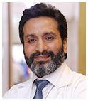
Dr. Susheel Bindroo

Director
Dr. Susheel Bindroo
Department
Pulmonary Medicine
Qualification
M.D, ID.C.C.M., F.N.B.(CCM), European Diploma in Intensive Care (EDIC), European Diploma in Adult Respiratory Medicine (EDARM), F.C.C.P. (USA),  

Interventional Pulmonology (Penn Medical Centre, Philadelphia)
Call us Call us on 022-61305000


Dr. Richa Mittal

Consultant
Dr. Richa Mittal
Department
Pulmonary Medicine
Qualification
  • DM (Pulmonary, Critical care and Sleep Medicine) - Gold Medal
  • MD, DNB (Pulmonary Medicine)
  • EDRM (London)
Call us Call us on 022-61305000


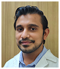
Dr. Pujan Parikh

Consultant
Dr. Pujan Parikh
Department
Pulmonary Medicine
Qualification
MBBS, MD (Pulmonary Medicine)
Call us Call us on 022-61305000


Download

RELIANCE FOUNDATION HOSPITAL Free Mobile App From

Google Play Icon
Get it onGoogle Play
App Store Icon
Download on theApp Store
  • Facebook Icon
  • X Icon
  • Youtube Icon
  • Instagram Icon

© 2024 Sir H. N. Reliance Foundation Hospital and Research Centre